
Wisdom teeth rarely announce themselves with much warning. One week things feel fine, and a few weeks later, there’s pressure, swelling, or pain in the back of the jaw. By the time symptoms appear, the problem has usually been developing for a while underneath the gumline, where nothing is visible from the outside.
This is one of the clearer cases where routine dental X-rays in Winnipeg genuinely change outcomes. They show what’s happening below the surface before symptoms develop, which affects the decisions available to the patient and the complexity of any treatment that becomes necessary.
What an Impacted Wisdom Tooth Actually Means
A wisdom tooth is considered impacted when it lacks enough space to emerge fully into its normal position in the arch. This can happen for a few reasons: the jaw may not be large enough to accommodate the tooth, the adjacent second molar may be blocking the path of eruption, or the tooth may be angled in a direction that prevents it from coming through the gum properly.
There are different degrees of impaction. A partially impacted tooth has broken through the gum partially, but can’t complete the eruption. A fully impacted tooth remains entirely below the gum and sometimes below the bone. The angle matters too: some impacted wisdom teeth are angled horizontally, directly toward the roots of the second molar. Others tilt forward, backward, or grow at an angle that puts pressure on adjacent structures without ever breaking through the gum.
Most people have four wisdom teeth, one in each corner of the mouth, but it’s not unusual to have fewer or, in some cases, none at all.
What X-Rays Show That a Clinical Exam Cannot
A standard dental examination can identify whether a wisdom tooth has partially erupted and whether the gum tissue around it looks healthy. What it cannot do is tell you the position of a tooth that’s still fully beneath the surface, how the roots are oriented, how close the tooth is to the nerve canal in the lower jaw, or whether an impacted tooth is putting pressure on the roots of the second molar next to it.
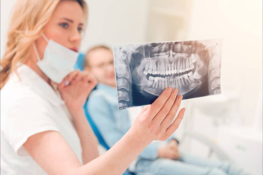
X-rays fill that gap. Periapical X-rays, which show individual teeth from crown to root tip, and panoramic X-rays, which capture the full upper and lower jaw in a single image, both provide information about the position, angle, depth, and root development of wisdom teeth that haven’t yet erupted.
At Linden Market Dental Centre, Dr. Morgan Winchar uses X-rays as part of routine dental exams to monitor wisdom tooth development over time. For younger patients, this often means tracking progression across several years of appointments, which produces a clearer picture of whether and how a wisdom tooth is likely to cause problems.
The Specific Problems X-Rays Catch Early
Pressure on adjacent molars
When a wisdom tooth is angled toward the second molar, its crown can press against the roots of that tooth as it continues to push forward. This pressure can cause root resorption, which is the gradual breakdown of the root structure in the affected tooth. In some cases, the damage to the second molar is significant enough to affect its long-term prognosis.
This process can be completely painless while it’s happening. X-rays are often the only way to identify it before the damage reaches a point where the second molar requires treatment of its own.
Cyst formation around impacted teeth
Fully impacted wisdom teeth are surrounded by a follicular sac, a small fluid-filled sac that’s a normal part of tooth development. In some cases, this sac can expand into a cyst, which enlarges within the jawbone and can displace adjacent teeth, weaken the bone, and, if large enough, cause structural problems.
Cysts associated with impacted teeth are typically asymptomatic in the early stages. Routine panoramic X-rays are how they’re discovered before they become significantly enlarged.
Infection risk from partial eruption
A partially erupted wisdom tooth creates a flap of gum tissue over part of the crown. This flap, called an operculum, traps food and bacteria in an area that’s almost impossible to clean properly with a toothbrush. The result is a condition called pericoronitis, an infection of the gum tissue around the partially erupted tooth.
Pericoronitis can cause swelling, pain, jaw stiffness, and in more severe cases, spreading infection. X-rays taken before the tooth fully erupts can show whether the angle and available space suggest that partial eruption is likely, which helps the dentist have an informed conversation with the patient about whether extraction makes sense before the pattern repeats.
When Dentists Typically Recommend Taking Wisdom Tooth X-Rays
Monitoring wisdom teeth with X-rays usually begins in the mid-to-late teenage years, often between 16 and 19, when root development is progressing and the likelihood of impaction can be assessed more accurately. At this stage, roots are typically not yet fully formed, which generally makes extraction less technically complicated if it does become necessary.
For patients in their twenties and beyond who haven’t had recent imaging, a panoramic X-ray taken at a new patient exam or a routine checkup will often reveal the current status of wisdom teeth that were never previously monitored.
Dr. Winchar reviews wisdom tooth position as part of dental exams at Linden Market Dental Centre and discusses findings and options with patients based on what the imaging shows.
Not Every Impacted Wisdom Tooth Needs to Be Extracted
This is worth stating clearly. Not all impacted wisdom teeth require immediate removal. If a wisdom tooth is fully impacted, positioned in a way that poses no risk to adjacent teeth, showing no signs of cyst formation, and not causing symptoms, some dentists and patients choose a monitoring approach rather than prophylactic extraction.
The decision depends on the specific anatomy shown on X-rays, the patient’s age, and the risk-benefit balance for that individual case. The value of having current X-rays is that the conversation can be based on what’s actually happening rather than on assumptions.
About Linden Market Dental Centre
Linden Market Dental Centre is located at 877 Waverley St, Unit 7, Winnipeg, MB, and serves patients across the Winnipeg community. Dr. Morgan Winchar graduated from the University of Manitoba Faculty of Dentistry and brings experience across general dentistry, restorative work, implants, and cosmetic care. The clinic accepts the Canadian Dental Care Plan (CDCP) and welcomes new patients.
Book a Dental Exam at Linden Market Dental Centre in Winnipeg
If you haven’t had recent X-rays and have questions about your wisdom teeth, or if you’re due for a routine exam, Linden Market Dental Centre is accepting new patients.
Call (204) 487-2226 to book your appointment.
- Book a dental exam with X-rays at Linden Market Dental Centre in Winnipeg
- Call us to ask about wisdom tooth assessment or monitoring
- Schedule an appointment with Dr. Winchar at our Waverley St location
- Contact Linden Market Dental Centre to ask about CDCP coverage for exams
- Request your new patient appointment at our Winnipeg dental clinic today.